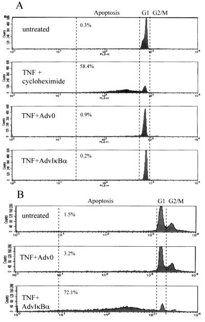
Figure 5.
Lack of apoptosis with TNFα in IκBα-infected macrophages. Human monocytes treated with M-CSF for 48 hr (A) or Hela cells (B) were plated on 60-mm Petri dishes at a density of 2–3 × 10⁶ cells per dish. They were left untreated or infected with control adenovirus or AdvIκBα at a moi of 40. Two days after infection, TNFα (20 ng/ml) and cycloheximide (2 μg/ml) were added as indicated. After 16 hr cells were stained for 30 min in 1.5 ml of a hypotonic fluorochrome solution (50 μg/ml of propidium iodide in 0.1% sodium citrate plus 0.1% Triton X-100), and the resulting propidium iodide-stained nuclei were analyzed by flow cytometry. The percentage of cells in each sector is indicated.

