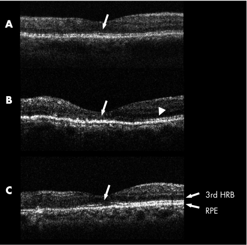
Figure 2 Optical coherence tomography (OCT) images of vertical retinal sections of the fovea obtained at the final visit. Macular oedema secondary to branch retinal vein occlusion (BRVO) is resolved completely in all images. The retina affected by BRVO is shown on the left in all images. Status of the third high reflectance band (HRB) was evaluated with monochromatic (greyscale) OCT images. (A) The third HRB is well preserved in the fovea (arrow). (B) The third HRB is well preserved in the unaffected retina (arrowhead), but is deteriorated in the fovea (arrow) and affected retina. (C) The third HRB is discontinuous in the fovea (arrow), while it is clearly detected in unaffected retina. RPE, retinal pigment epithelium.

An official website of the United States government
Here's how you know
Official websites use .gov
A
.gov website belongs to an official
government organization in the United States.
Secure .gov websites use HTTPS
A lock (
) or https:// means you've safely
connected to the .gov website. Share sensitive
information only on official, secure websites.
