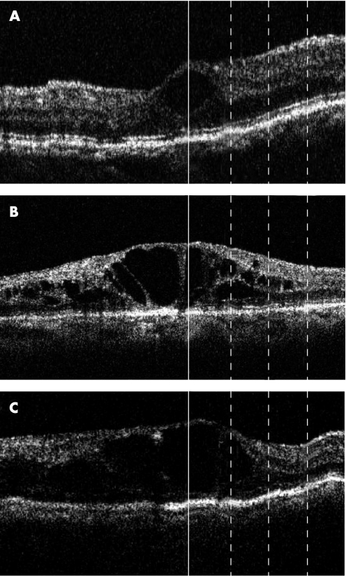
Figure 3 Monochromatic (greyscale) images of optical coherence tomography (OCT) obtained at the initial visit. All images show macular oedema secondary to branch retinal vein occlusion (BRVO). The retina affected by BRVO is on the left in all images. The vertical line at the centre of each image represents the location of the fovea. The three dotted lines represent points at 500, 1000 and 1500 µm from the fovea, from the left, respectively. (A) The third high reflectance band (HRB) is clearly visible at all points. (B) The third HRB is not detected beneath the fovea or at 500 µm from the fovea, but does appear at the 1000 µm and 1500 µm points. (C) The third HRB can be detected only at 1500 µm from the fovea.

An official website of the United States government
Here's how you know
Official websites use .gov
A
.gov website belongs to an official
government organization in the United States.
Secure .gov websites use HTTPS
A lock (
) or https:// means you've safely
connected to the .gov website. Share sensitive
information only on official, secure websites.
