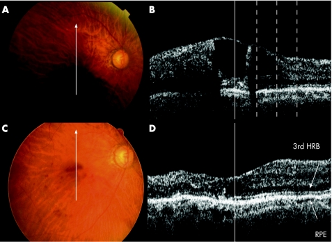
Figure 4 An 82‐year‐old woman with branch retinal vein occlusion (BRVO) accompanied by macular oedema (MO) with a 1‐month history of decreased visual acuity in the right eye, which was 20/50 at the initial visit. (A) Fundus photograph shows extensive retinal haemorrhage associated with BRVO. (B) Monochromatic optical coherence tomography (OCT) image of the fovea was made at the initial visit. The vertical solid line represents the location of the fovea. The three dotted lines represent points 500, 1000 and 1500 µm from the fovea, from the left, respectively. MO was prominent with large cystoid spaces and a foveal thickness of 464 µm. The third HRB cannot be detected in the fovea but is well visualised at other measurement points. The patient was treated with grid laser photocoagulation in the right eye. (C) Fundus photograph at the final visit shows minimal retinal haemorrhage. (D) OCT image at the final visit shows the MO to be completely resolved. Foveal thickness is now 165 µm. The third HRB is well preserved in the fovea and visual acuity was 20/25. The vertical solid line represents the location of the fovea.

An official website of the United States government
Here's how you know
Official websites use .gov
A
.gov website belongs to an official
government organization in the United States.
Secure .gov websites use HTTPS
A lock (
) or https:// means you've safely
connected to the .gov website. Share sensitive
information only on official, secure websites.
