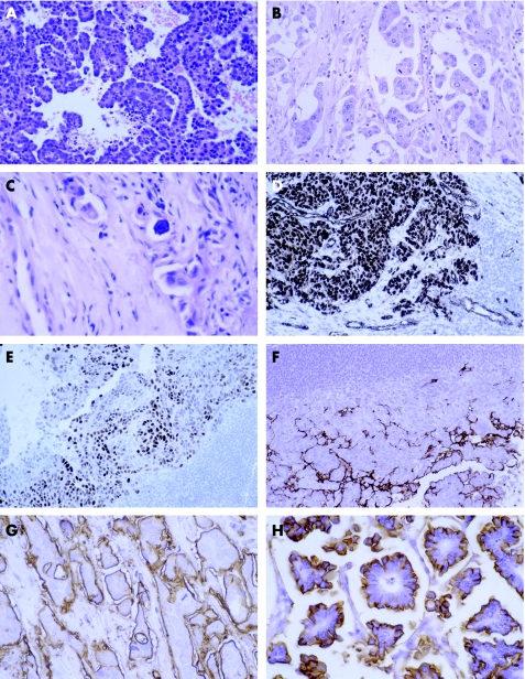
Figure 1 Metastasis from serous papillary carcinoma of the ovary: (A) typical papillary architecture; (B) less typical papillary architecture and (C) calcification. Immunohistochemical analysis shows expression of (D) Wilms' tumour 1 in tumour nuclei and vessels, (E) oestrogen receptor and (F) Ca125. Epithelial membrane antigen expression in (G) metastasis from serous papillary carcinoma of the ovary compared with (H) invasive micropapillary carcinoma of the breast.

An official website of the United States government
Here's how you know
Official websites use .gov
A
.gov website belongs to an official
government organization in the United States.
Secure .gov websites use HTTPS
A lock (
) or https:// means you've safely
connected to the .gov website. Share sensitive
information only on official, secure websites.
