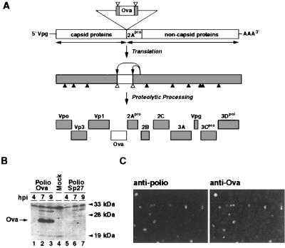
Figure 1.
Expression and stability of recombinant poliovirus expressing Ova. (A) Schematic diagram of a recombinant poliovirus Polio-Ova and strategy for expression of Ova. The bar represents recombinant poliovirus genomic RNA. The sequence of the C-terminal half of Ova (empty bar) was inserted at the junction between the coding regions for capsid and noncapsid proteins. The Ova sequence was flanked by cleavage sites for the viral protease 2Apro. After translation, the viral polyprotein is proteolytically processed, resulting in the release of the foreign peptide and the generation of mature and functional viral proteins. ▴ indicate 3Cpro cleavage sites, ▵ represent 2Apro cleavage sites. (B) Expression of the exogenous protein in cells infected with the recombinant virus Polio-Ova. Cytoplasmic lysates from HeLa cells infected with Polio-Ova or Polio-Sp27 recombinant viruses were analyzed by Western blot with antibodies directed against Ova. Cytoplasmic lysates were prepared after 4 (lanes 1 and 5), 7 (lanes 2 and 6), and 9 hr (lanes 3 and 7) postinfection (hpi). Lanes 1–3, extracts from Polio-Ova-infected HeLa cells; lanes 5–7, Polio-Sp27-infected cells; and lane 4, mock-infected cells. Molecular weight markers indicate relative mobility. Specific bands detected by antibodies directed against the Ova are indicated by an arrow. (C) Analysis of the stability of the recombinant poliovirus Polio-Ova by immunofluorescence. HeLa cells were infected at low MOI (<1) with Polio-Ova obtained after two successive passages in HeLa cells. Five hours postinfection, cells were fixed with 2% paraformaldehyde and stained with a mAb directed against poliovirus 2C protein and a polyclonal rabbit antibody that reacts with Ova. A portion of the entire field studied is shown. More than 200 infected cells were examined to determine the proportion of revertants in the viral stock.

