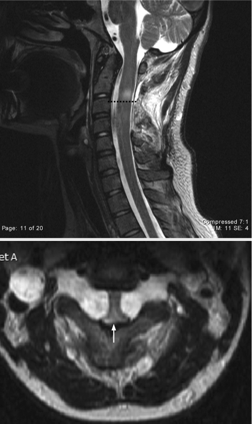
Figure 1 T2 weighted sagittal (top) and axial (bottom) MRI of the upper cervical spine showed a 1–2 mm reduction in the size of the spinal canal (arrow) in a 17‐year‐old female (case No 1) who presented with progressive quadriparesis, hyperreflexia and up‐going toes. The broken line denotes abnormal signal intensity in the area of cord compression.

An official website of the United States government
Here's how you know
Official websites use .gov
A
.gov website belongs to an official
government organization in the United States.
Secure .gov websites use HTTPS
A lock (
) or https:// means you've safely
connected to the .gov website. Share sensitive
information only on official, secure websites.
