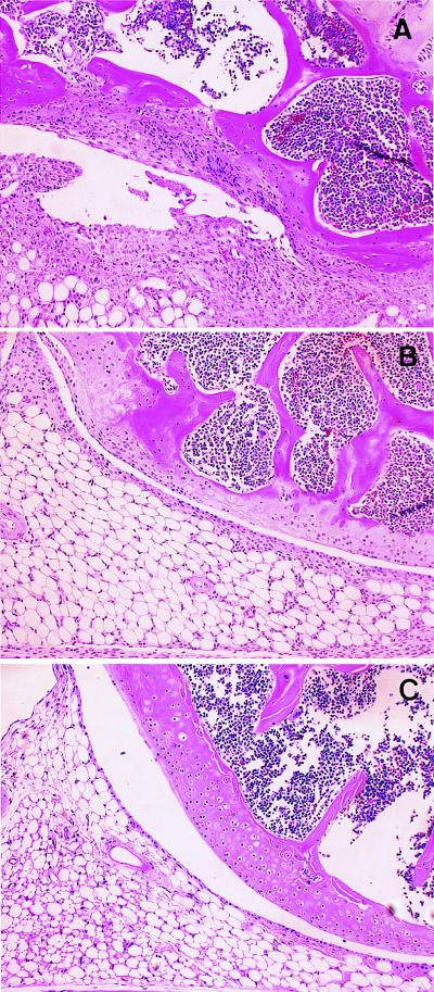
Figure 1.
Representative histopathologies of the knee joints stained with hematoxylin and eosin (×100). Thirty-five days after the first immunization, mice were sacrificed and subjected to histopathological examination. (A) Wild-type (IL-6 +/+) mice induced AIA (grade 4); massive cellular infiltration, synovial hyperplasia, neovasculization, and erosion of cartilage and bone were observed in the knee joint. (B) IL-6-deficient (IL-6 −/−) mice (grade 2); only limited synovial lining cell hyperplasia was detected. (C) Wild-type (IL-6 +/+) mice injected with saline as a control (grade 0); no features of synovitis were detected.

