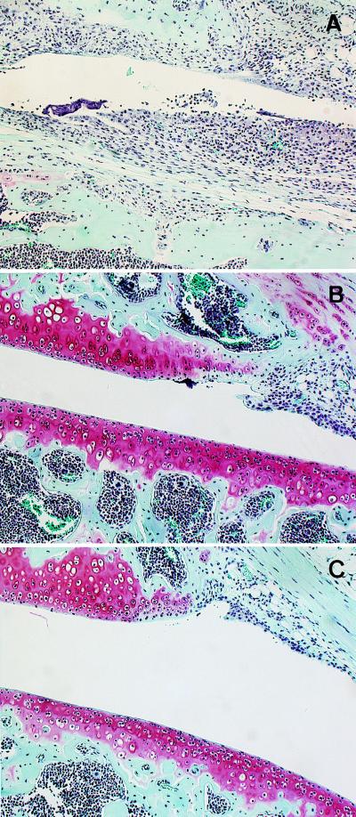
Figure 2.
Femoropatellar sections of the knee joints stained with Safranin O/fast green (×100). (A) Wild-type (IL-6 +/+) mice induced AIA; severe synovitis with pannus formation in patella (upper side) and femur (lower side) was observed. Moreover, Safranin O was not stained, indicating complete cartilage destruction. (B) IL-6-deficient (IL-6 −/−) mice induced AIA; only limited synovial hyperplasia was detected. Safranin O was stained strongly in patella and femur (red color), indicating cartilage was completely preserved. (C) Wild-type (IL-6 +/+) mice injected saline as a control; no features of synovitis were detected, and Safranin O was strongly stained.

