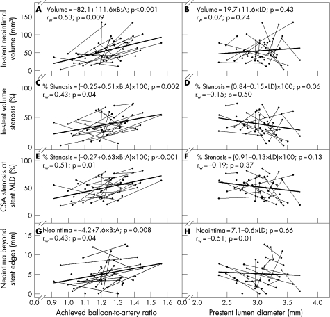
Figure 4 Primary outcome variables. Balloon‐to‐artery (B:A) ratio and prestent lumen diameter (LD) plotted against in‐stent neointimal volume (A, B), in‐stent volume stenosis (C, D), cross‐sectional area (CSA) stenosis at the stent minimum lumen diameter (MLD) (E, F) and neointima beyond the stent edges (G, H). Data are from 49 vessels in 27 animals that survived to day 28 and had evaluable intravascular ultrasound images. Thin lines connect data points (solid circles) from the same animal. Data points with no connecting line are from five animals that received a single stent. Linear regression lines (thick lines), linear regression equations and p values for test of slope (first p value in each panel) are based on multilevel regression analysis, which takes into account non‐independence of data obtained from multiple vessels within the same animal. A test‐of‐slope p value <0.05 indicates that the slope of the regression line is significantly different from zero. Within‐animal correlations (rw) and their associated p values are based on data from 22 animals that underwent stent placement in two vessels. A within‐animal correlation p value <0.05 indicates that there is a significant association between the x‐axis and y‐axis variables within the animal. A larger B:A ratio was associated with greater in‐stent neointimal volume (A), in‐stent volume stenosis (C), CSA stenosis at the stent MLD (E) and neointima beyond the stent edges (G). The regression equation in C predicts a 31% in‐stent volume stenosis at a B:A ratio of 1.1:1. The regression equation in E predicts a 42% CSA stenosis at the stent minimum lumen diameter at a B:A ratio of 1.1:1. Note that in H, prestent LD was not significantly associated with neointima beyond the stent edges using multilevel regression analysis, but there was a significant negative within‐animal correlation; within an individual animal, larger prestent LD was associated with less neointima beyond the stent edges. This finding shows that data points obtained from multiple vessels within the same animal cannot be assumed to be independent of each other.

An official website of the United States government
Here's how you know
Official websites use .gov
A
.gov website belongs to an official
government organization in the United States.
Secure .gov websites use HTTPS
A lock (
) or https:// means you've safely
connected to the .gov website. Share sensitive
information only on official, secure websites.
