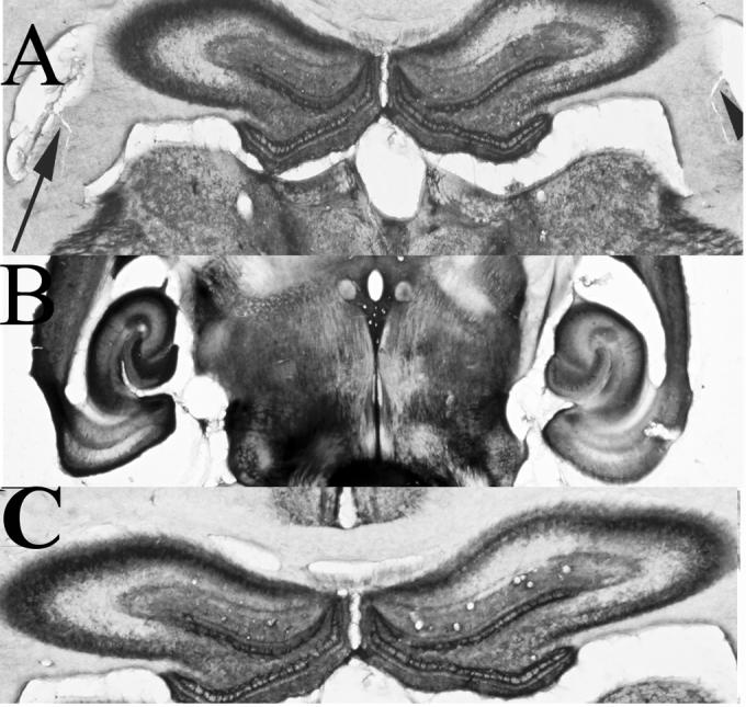
Figure 6. Experiment 2 Acetylcholinesterase.

A. Acetylcholinesterase stain of rodent hippocampus showing continued presence of AChE as well as fimbria transection. Arrowheads point to bilateral fimbria transection. B. Ventral hippocampus stained for AChE after fimbria transection. C. Control brain processed for AChE. Note there were no differences between the control and transected brains with respect to AChE staining.
