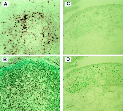
Figure 1.
B19 DNA and VP-1 in synovial specimens. (A) ISH of RA synovium shows nuclear stain for B19 DNA and RNA (purple) in the cells mainly in lymphoid follicle. (B) Stain for anti-VP-1 antibody. The composed cells in the germinal center of lymphoid follicle and the mononuclear cells infiltrated in the sublining layer are stained on the RA synovium (brown). (C) The composed cells of OA synovium are negative for B19 DNA and RNA, when determined by ISH. (D) Stain for anti-VP-1 antibody. The SVC are not stained for anti-VP-1 antibody on OA synovium. All original magnifications ×400.

