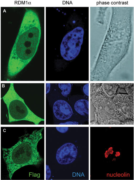
Figure 2.
Subcellular localization of RDM1α. (A–B) Confocal microscopic images of HeLa cells expressing EGFP-RDM1α (A) and HEK293T cells expressing RDM1α-EGFP (B). Cells were fixed and counterstained with DRAQ5 (blue) prior to visualization. Green represents EGFP-tagged RDM1α. (C) HEK293T cells transfected with pFlag-RDM1α were double stained for nucleolin (red) and the Flag epitope (green). Cell transfection and immunofluorescence analysis were carried out as described under ‘Materials and methods’ section.

