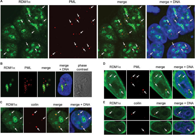
Figure 4.
Association of RDM1α-EGFP with PML and CBs. (A) Confocal micrographs of RDM1α-EGFP-transfected HEK293T cells following treatment with MG132 (10 μM, 4 h) and staining of PML (red). The DNA was counterstained with DRAQ5 (blue). In addition to accumulating in the nucleoli, RDM1α can be seen in numerous dot-like nuclear structures. Several of these dots (some of which are indicated by arrows) coincide with or are in close proximity to PML bodies as illustrated in the merged image. Note that, in the cells depicted here, no PML protein is detected in the nucleoli colonized by RDM1α. (B) Image of a MG132-treated cell (10 μM, 8 h) showing the partial accumulation of PML protein in nucleoli colonized by RDM1α. (C) HEK293T cells transfected with RDM1α-EGFP were treated with MG132 (10 μM, 2 h), followed by staining of the CB marker coilin (red). As illustrated in the merged image, some of the dot-like structures containing RDM1α (indicated by arrows) colocalize, or are in close contact with CBs in MG132-treated cells. (D–E) Association of RDM1α with PML bodies (D) and CBs (E) in unstressed cells. The panels in (E) represent two sections of the same cell showing dot-like structures containing RDM1α (indicated by arrows) that colocalize with CBs.

