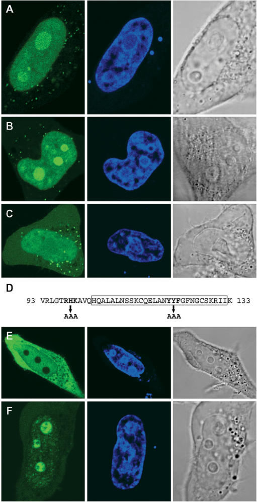
Figure 7.
Nuclear and nucleolar localization signals in exon 3 of RDM1α. (A–C) HeLa cells expressing either wt exon 3 (A), the (120–122) YYF-AAA variant (B) or the (98–100) RHK-AAA variant (C) as EGFP-RDM190–133 fusion proteins were fixed and observed by direct fluorescence, under a confocal microscope. (D) Amino-acid sequence of human RDM1 exon 3, showing the position of the mutations carried out as well as the RD motif (boxed). (E–F) Deletion of exon 3 does not prevent the nucleolar accumulation of RDM1α in response to heat stress. HeLa cells expressing EGFP-RDM1α-ΔE3 were observed in the absence of stress (E) or following mild heat-shock treatment (43°C, 30 min) (F). Green represents EGFP-tagged exon 3 or RDM1α-ΔE3 and blue represents DRAQ5-stained nuclei. See text for details.

