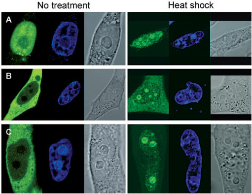
Figure 8.
Subcellular localization of the RRM domain as well as N-terminal truncation mutants of RDM1α. Confocal microscopic visualization of control (left panel) or heat-shocked (right panel) HeLa cells expressing EGFP-RDM11–92 (A), EGFP-RDM190–284 (B) or EGFP-RDM1133–284 (C). Cells were fixed and counterstained with DRAQ5 (blue) prior to visualization.

