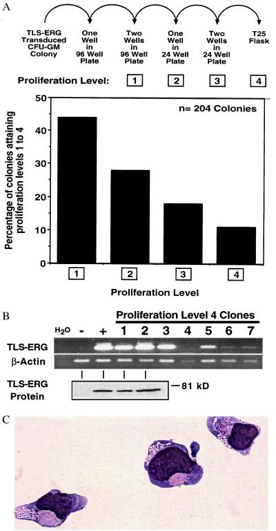
Figure 4.
TLS-ERG-transduced CFU-GM colonies proliferate in liquid culture. (A, upper) TLS-ERG-transduced G418-resistant CFU-GM colonies (from six independent experiments) were placed in liquid culture containing 300 ng/ml SCF, 300 ng/ml GM-CSF, and 50 ng/ml IL-3 and allowed to progress through 4 proliferation levels in the absence of G418. (Lower) Bar graph depicting the percentage of 204 colonies attaining a given proliferation level. (B) RT-PCR (Upper) and Western blot (Lower) analysis revealing the presence of TLS-ERG transcripts and protein in nonproliferating level 4 clones, respectively. Although H2O represents the reagent control for RT-PCR, the negative (−) and positive (+) controls for RT-PCR and Western blot analyses are from PG13MSCVNEO and PG13MSCVTLS-ERG retroviral producer cells, respectively. (C) May–Grünwald–Giemsa stained cytospin preparation of TLS-ERG-transduced CFU-GM-derived cells that had been proliferating in liquid culture for 3 weeks.

