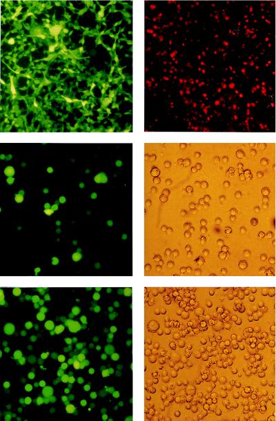
Figure 4.
(Top Left) GFP expression in 293 B95.8/F factor-positive cell lines. Living cells were examined with an inverse microscope under UV light. In both cases, the bright green color of all cells demonstrates a high level of expression of the GFP gene, which is part of the F factor backbone, as shown schematically in Fig. 1. (Top Right) Expression of the viral capsid antigen (VCA) in 293 carrying the B95.8/F factor DNA after induction of the lytic cycle. Fixed cells were incubated with an antibody against VCA and a second anti-mouse antibody coupled to the Cy5 fluorochrome. Stained cells were exposed to UV light. (Middle) GFP expression in cells incubated with supernatants from induced 293 cell lines carrying the B95.8/F factor DNA. Raji cells (1 × 10⁵) were incubated with 0.5 ml of supernatant from BZLF1-transfected 293 cells carrying the B95.8/F factor. Approximately 50% of the cells were GFP-positive, indicating a virus titer of at least 105 infectious viruses per ml. GFP fluorescence was investigated 48 hr after infection (Left). (Middle Right) Phase-contrast light microscopy. (Bottom) Primary human B cells were infected with supernatants from BZLF1 transfected 293 cells carrying the B95.8/F factor. As a consequence, immortalized B cell lines were generated that were investigated for GFP expression about 6 weeks after infection. (Bottom Left) UV light exposure. (Bottom Right) Phase-contrast light microscopy.

