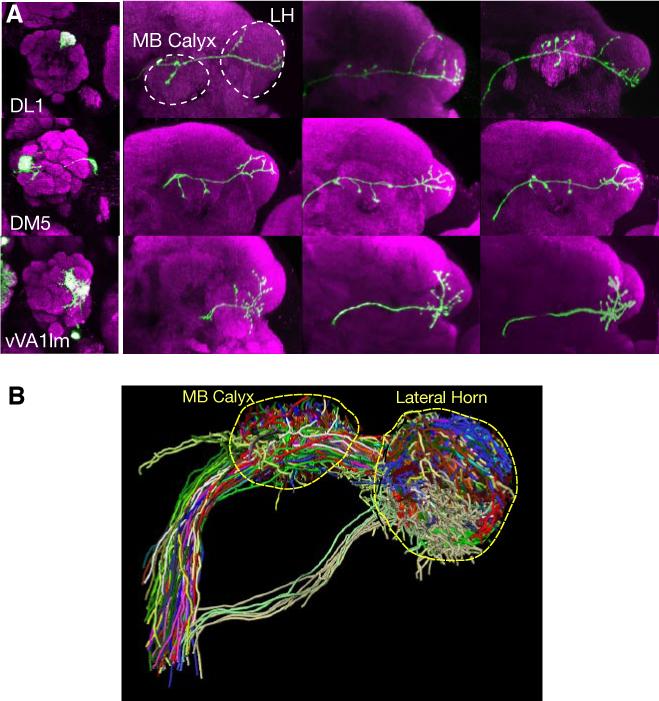
Fig. 2.

Analysis of MARCM-labeled single olfactory projection neurons revealed organization of higher olfactory centers with respect to input channels. (A) Representative images of PN axon arborization in the mushroom body and lateral horn from 3 individual animals are shown (right) for three different classes of PNs. PNs are classified based on glomeruli of the targeted dendrites as indicated (left). MB: mushroom body; LH: lateral horn. Modified after Marin et al. (2002). (B) MARCM labeled single cells are projected onto a standard brain after image registration (Jefferis et al., 2007). Shown are 28 classes of PNs (each labeled by a different color), with at least 2 example neurons shown per class.
