
Figure 5.
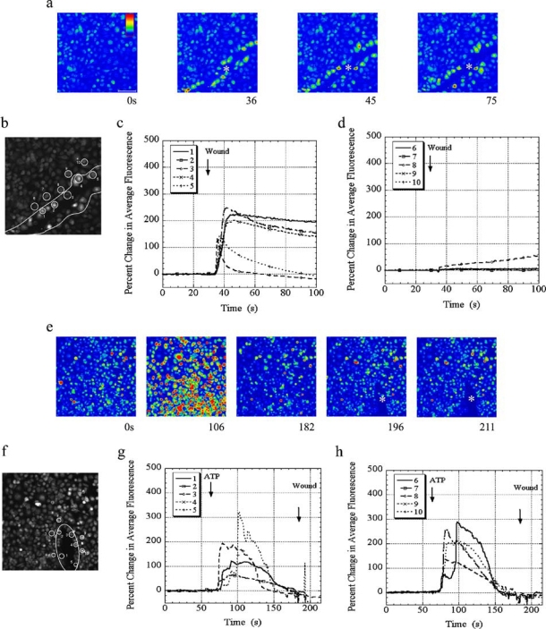
Ca2+ free media inhibits an injury response after ATP stimulation. Primary corneal epithelial cells were incubated in 5 µM Fluo-3AM for 30 min and imaged in a flow through apparatus on an LSM 510 confocal. Cells were washed in HEPES buffered saline for at least 30 s, and stimulated by wounding or ATP. Individual cells were analyzed for percent change in average fluorescence. Intensity scale is shown in (a) with red indicating highest Ca2+ levels and blue indicating lowest Ca2+ levels. The horizontal white bar in (a) represents 100 µm. a) Cells were pre incubated in BAPTA (100 µM) washed in HEPES buffer containing Ca2+ and wounded. A series of images taken from a time course of a wound (shown at asterisk) of a representative experiment is presented. b) A single image taken after wounding is shown. Cells (#1–5) immediately adjacent to the wound (c) and cells away (#6–10) from the wound (d) were analyzed. e) Cells were washed in Ca2+ free HEPES buffer containing EGTA, stimulated with 100 µM ATP, and wounded. A series of images taken from a representative time course is presented (wound shown at asterisk). f) A single image taken after wounding is shown. Cells (#1–5) immediately adjacent to the wound (g) and cells away (#6–10) from the wound (h) were analyzed. Images are representative of at least 10 independent experiments. The series of images are taken from Movie 2 (see online version of article at www.springeronline.com).