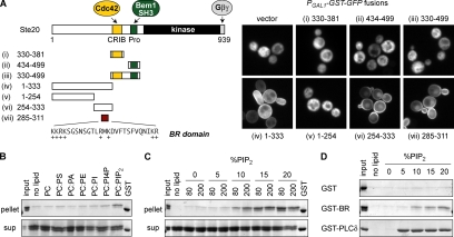
Figure 1.
The Ste20 regulatory region contains a membrane-binding domain. (A) Left, fragments used to map a membrane-targeting domain in the Ste20 N-terminus. Fragment 330-381 spans the minimal Cdc42-binding motif predicted from prior studies (Kim et al., 2000; Morreale et al., 2000; Gladfelter et al., 2001), although it is part of a larger conserved region that also includes the autoinhibitory domain (see Supplementary Figure S1). Right, Ste20 fragments were expressed as GST-GFP fusions (pPP1843, pPP1878, pPP1880, pPP1961, pPP1877, pPP1939, pPP1940, or pPP2428) from a galactose-inducible promoter in wild-type cells (PPY1368). A minimum region for membrane localization maps to residues 285-311, denoted the basic-rich (BR) domain. (B) The Ste20 BR domain binds liposomes containing PIP2. A purified GST-BR fusion (Ste20 residues 285-311) was mixed with 80 nmol of sucrose laden liposomes containing phosphatidylcholine (PC) alone or 80% PC plus 20% (mol/mol) of phosphatidylserine (PS), phosphatidic acid (PA), phosphatidylethanolamine (PE), phosphatidylinositol (PI), phosphatidylinositol-4-phosphate (PI4P), or PIP2. Liposomes were pelleted, and protein in bound (pellet) and unbound (sup) fractions is shown. “Input” shows 25% of input protein (=0.5 μg). GST was loaded on each gel as a size marker. (C) Dependence of GST-BR binding on concentration and density of PIP2. Binding assays (in 200 μl volume) used 80 or 200 nmol of PC liposomes with varying molar percentage of PIP2 (0–20%). “Input” shows 10% of input protein (=0.2 μg). (D) Comparison of liposome binding by GST alone, GST-BR, and GST-PLCδPH, using 80 nmol of PC-based liposomes with 0–20% PIP2. Only pellet fractions are shown. “Input” shows 12.5% of input protein (=0.25 μg).

