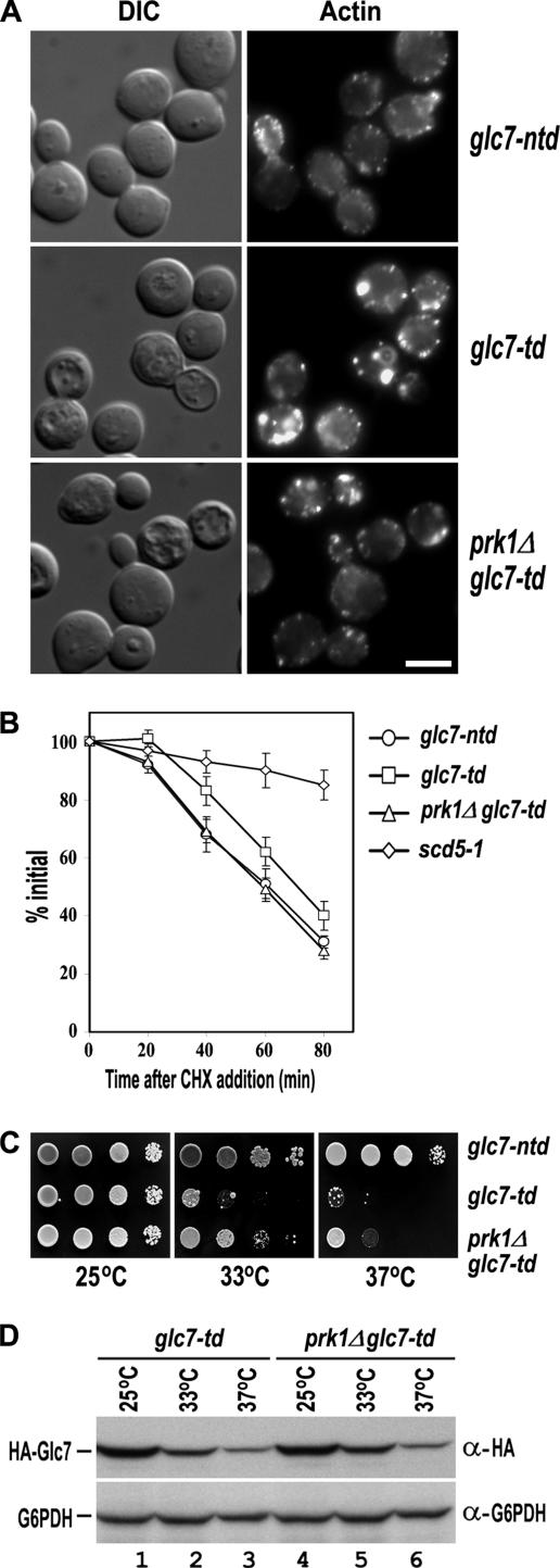
Figure 4.
Suppression of glc7-td by prk1Δ. (A) Actin staining of glc7-ntd (YMC499), glc7-td (YMC500), and prk1Δ glc7-td (YMC501) cells. The strains were grown at 25°C to early log phase and incubated at 37°C for 6 h followed by fixation and staining with rhodamine-phalloidin. Bar, 4 μm. (B) Uracil permease internalization assay of glc7-ntd, glc7-td, prk1Δ glc7-td, and scd5-1 cells. The cells were transformed with pFUR4-424, and the transformants were grown at 25°C to OD600 of 0.2–0.3. After incubated at 37°C for 6 h, cultures were brought back to 25°C, and the internalization of uracil permease was initiated by the addition of cycloheximide (CHX). Transport of [3H]uracil into yeast cells was used as a measurement for the relative amount of uracil permease retained on the cell surface. Uracil uptake is plotted as the percentage activity relative to the initial time point. The results shown are the averages of two independent assays. (C) The growth of glc7-ntd, glc7-td, and prk1Δ glc7-td strains at different temperatures. Cells were grown at 25°C to OD600 of 0.5, and 10× serial dilutions of each culture were dropped onto CuSO4-containing plates for incubation at the indicated temperatures for 2 d. (D) Comparison of the Glc7p protein level in glc7-td, and prk1Δ glc7-td strains. Cells were grown at 25°C to log phase and then shifted to either 33°C or 37°C for 1 h. Samples were taken at 0 and 60 min. Total proteins of each sample were prepared and subjected to Western analysis.

