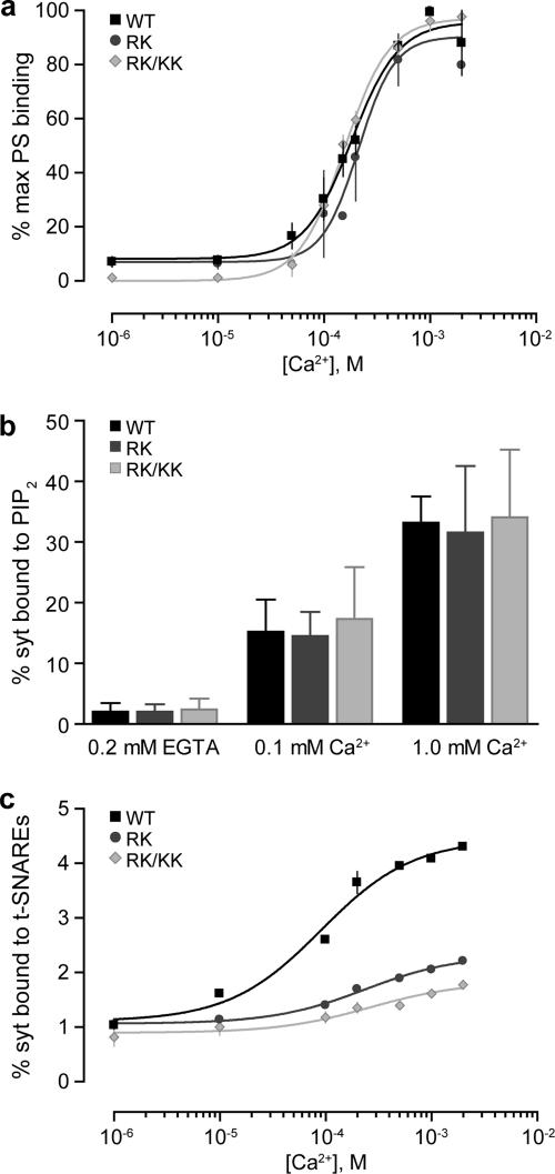
Figure 4.
C2A loop 2 and C2B loop 1 mutants exhibit decreased Ca2+-dependent SNARE binding but normal Ca2+-dependent phosphatidylserine binding. (a) C2AB, C2AB(R199A/K200A) (RK) or C2AB(R199A/K200A/K297A/K301A) (RK/KK) were incubated with protein-free PC liposomes containing 15% PS at the indicated Ca2+ concentrations and bound C2AB was determined as in Figure 3b. Maximal bound C2AB was set equal to 100% and plotted (±SE, n = 3) as a function of [Ca2+]. (b) C2AB, C2AB(R199A/K200A) (RK), or C2AB(R199A/K200A/K297A/K301A) (RK/KK) were incubated with protein-free PC liposomes containing 5% PIP2 at the indicated Ca2+ concentrations and bound C2AB was determined as in Figure 3b. Fractional bound C2AB was plotted (±SE, n = 3) as a function of [Ca2+]. (c) C2AB and indicated mutants were incubated with PC liposomes containing SNARE complexes at the indicated Ca2+ concentrations, and bound C2AB was determined as in Figure 3b. The fraction of C2AB bound was plotted (±SE, n = 3) as a function of [Ca2+].

