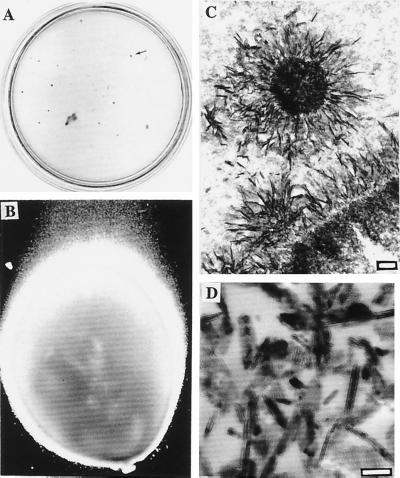
Figure 2.
Nanobacterial stony colonies and comparison to hydroxyapatite. (A) Colonies on modified Loeffler medium in a 10-cm plate. The colonies penetrated through the medium forming stony pillars. Arrow shows one typical grayish brown colony depicted in B. (×40.) (C) Needle-like crystal deposits in the pillar revealed by TEM. (Bar = 200 nm.) (D) TEM image of reference apatite crystals. (Bar = 100 nm.)

