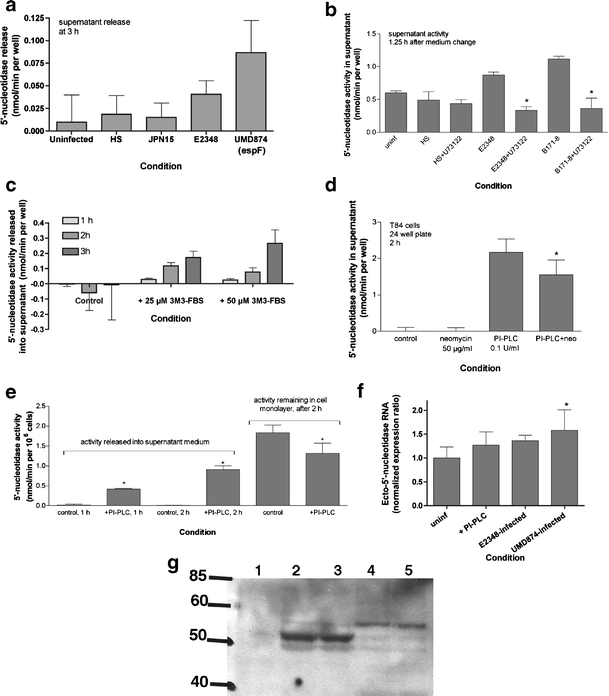
Fig. 3.

a Effect of EPEC mutants and PI-PLC inhibitors and activators on induced 5′-nucleotidase release into supernatant. Release of 5′-nucleotidase activity into supernatant medium was measured as described in “Materials and methods” and in the legend to Fig. 2. Once again, activity in the cell-free sterile filtrates is expressed as nmol/min per well. However, for purposes of comparison 48-well plates contain ~0.25 × 10⁶ T84 cells per well at confluency. b U73122, an inhibitor of PI-PLC, was used at a concentration of 2 μm and was re-added after the medium change; *significantly decreased compared to the EPEC strain alone, p < 0.05. cm-3M3-FBS, a cell-permeant sulfonamide activator of PI-PLC, was added at the concentrations and for the times indicated. d Inhibitory effect of neomycin on PI-PLC-induced 5′-nucleotidase release; this experiment was performed on cells grown in a 24-well plate, with ~0.8 × 10⁶ cells per well; *significantly decreased compared to PI-PLC alone. e PI-PLC was again added to a final concentration of 0.1 U/ml for the times shown; *significantly different from the corresponding control. This figure is a composite of experiments that were separated in time and with cells of different passage number; therefore the absolute amount of activity varies among the figure parts. f Effect of EPEC infection on expression of RNA encoding ecto-5′-nucleotidase in T84 cells, by reverse transcription and real-time PCR. T84 cells were infected for 35 min, then the supernatant medium was changed to remove unattached bacteria, then the T84 cell monolayer harvested for RNA extraction 4 h after the medium change. Reverse transcription and PCR conditions were as described in “Materials and methods.” Expression of ecto-5′-nucleotidase was normalized to that of glyceraldehyde 3-phosphate dehydrogenase (GAPDH). f Normalized expression of a single representative experiment (mean ± SD of 6 PCR wells). g Immunoblot analysis of the proteins released into the supernatant after 2 h of infection with EPEC or an EPEC mutant (SE1010, sepZ/espZ), using monoclonal antibody against CD73. Lane 1, supernatant from uninfected, control T84 cells. Lanes 2 and 3, supernatant from cells infected with wild-type E2348/69. Lanes 4 and 5, supernatants from cells infected with the sepZ mutant. GPI-linked CD73 has an apparent molecular size of 72 kDa (not seen in this blot) and the GPI-cleaved soluble portion runs at 51 kDa (heavy band in lanes 2 and 3)
