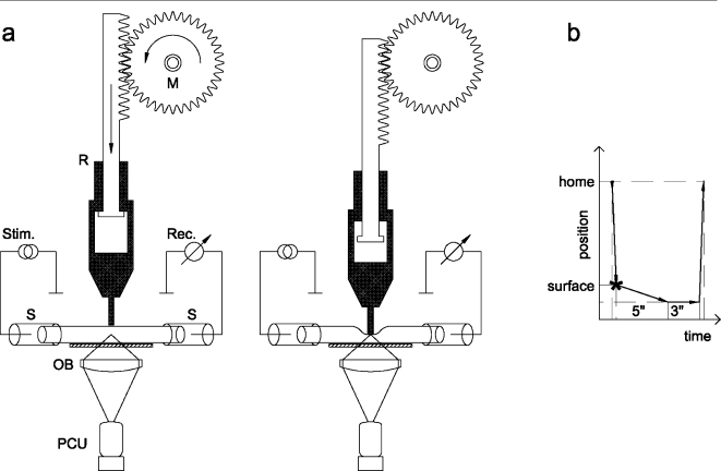
Figure 2.
Schematic drawing of the method applied for detection of ATP release during compression of isolated spinal roots. (a) A compression rod attached to a motorized micromanipulator was positioned perpendicular to the tissue and was brought to a point above the surface of an isolated spinal root. The rod consisted of two parts, the lower part was free to move against the upper part. The end of the rod had a diameter of 1 mm; the width of a thick dorsal spinal root is about 0.8 mm. The rod was advanced slowly by means of the manipulator motor. The bath solution contained an ATP assay mix; bioluminescence was detected by the photon counting unit (PCU). In addition, suction electrodes at both ends of the isolated spinal roots were used for stimulation and recording of compound action potentials. (b) Illustration of the cycle of nerve compression: the rod moved from the home position above the spinal root to the tissue surface, reached its lowest position within 5 s, halted at this position for 3 s, and moved upward afterwards to the home position. The total time of the compression period was about 8 s

