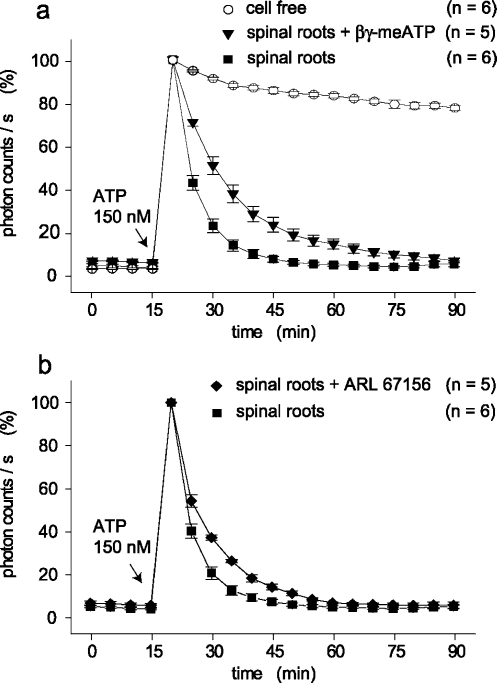
Figure 6.
Modulation of ATP degradation by ecto-ATPase inhibitors. Kinetics of ATP degradation were tested in culture dishes which contained two pairs of spinal roots each. The concentration of ATP was measured every 5 min and is given in photon counts/s relative to the light intensity at 150 nM ATP. This concentration of ATP was added 15 min after the start of the measurements and degradation of ATP was followed for a further 75 min. (a) A comparison was made between cell-free culture dishes, spinal roots, and spinal roots in the presence of βγ-meATP (300 µM). Degradation of ATP is much faster in the presence of spinal roots as compared to ATP consumption by a solution containing the ATP assay mix only; βγ-meATP had an inhibitory effect on ATP hydrolysis by spinal roots which is statistically significant (see text). (b) Kinetics of ATP degradation was tested after addition of ARL 67156 (100 µM). Also this compound slightly, but significantly (see text), reduced hydrolysis of ATP seen in the presence of spinal roots

