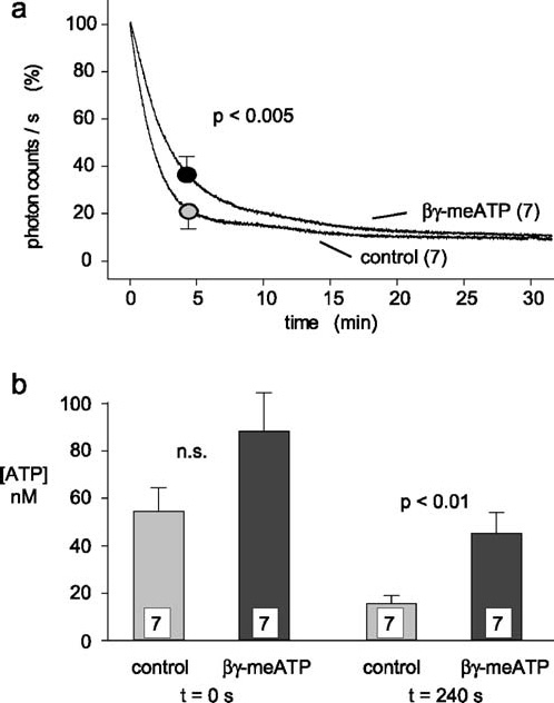
Figure 8.

Effects of βγ-meATP on compression-induced release of ATP. Fourteen different isolated spinal roots were tested with the experimental protocol illustrated in Figure 7. (a) Averaged data of the decline in light production observed after compression of isolated spinal roots tested either in control solution or in the presence of βγ-meATP (300 µM). The maximal light production (in photon counts/s) was set to 100% and data obtained after 4 min were used for statistical analysis. Note that βγ-meATP produces a small, but significant, slowing of the decay time. (b) Averaged data from the experiments illustrated in (a) are plotted as concentrations of ATP reached in the bath solution immediately after nerve compression (t = 0 s) and 4 min later (t = 240 s). Compression of spinal roots in the presence of βγ-meATP resulted in higher concentration of ATP and statistically significant slower recovery
