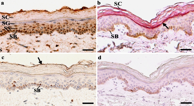
Figure 1.
Expression of P2X5 and P2X7 receptors in paraffin sections of normal human skin. Nuclei were counterstained blue with haematoxylin. (a) In normal skin, P2X5 immunoreactivity (brown) was found in basal keratinocytes (B) (although this was not easy to distinguish from melanin), and in the stratum spinosum (SS), and in very few cells in the stratum granulosum (SG). P2X5 receptor staining was absent from the stratum corneum (SC), apart from at the outer edge. P2X5 receptor staining was confined largely to the cell membranes in the basal layer, and found in the cytoplasm, and occasionally in the nucleus in both basal and suprabasal keratinocytes. Scale bar. 25 μm. (b) P2X7 immunoreactivity (pink) was present in the epidermis of all normal skin samples, and was associated with cells and cell fragments (arrow) in the stratum corneum (SC). Note the brown melanin in the basal layer (B). Scale bar, 25 μm. (c) There was residual staining of the outermost edge of the stratum corneum (arrow) with the P2X5 receptor antibody no primary control, and therefore this was non-specific staining. Note the brown melanin in the basal layer (B). Scale bar, 25 μm. (d) There was no staining in the no primary control for the P2X7 receptor antibody Scale bar, 25 μm.

