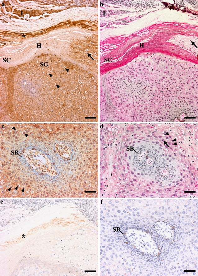
Figure 2.
Expression of P2X5 and P2X7 receptors in paraffin sections of human warts. Nuclei were counterstained blue with haematoxylin. (a) Low power view of P2X5 immunoreactivity (brown) in the wart. P2X5 receptors were present within the keratinocytes of the wart. There was marked hyperkeratosis (H), which was negative for P2X5 receptors, although areas of parakeratosis were positive (arrow). At the outer edge of the stratum corneum there was a band of heavy staining (asterisk). P2X5 receptors were also found in the inflammatory cell infiltrate (I) above the stratum corneum (SC). There was a prominent granular layer (SG), within which cells (koilocytes) showed typical cytoplasmic vacuolation (arrowheads). Scale bar. 100 μm. (b) Low power view of P2X7 immunoreactivity (pink) in the wart. P2X7 receptors were strongly present within the hyperkeratotic (H) areas of the stratum corneum (SC), but not in areas of parakeratosis (arrow). P2X7 receptors were weakly present in the wart keratinocytes, and mainly found in the nucleus. P2X7 receptors were also weakly found in the inflammatory cell infiltrate (I) above the stratum corneum. Scale bar, 100 μm. (c) High power view of P2X5 immunoreactivity (brown) in wart keratinocytes. There were a few positive cells in the basal layer (B), but most of the positively stained cells were in the suprabasal layers. Koilocytes showed P2X5 receptor staining in the nucleus (arrowheads). Scale bar, 50 μm. (d) P2X7 immunoreactivity (pink) was present in the suprabasal layers of the wart, in either large, flat nuclei with an obvious nuclear membrane (arrow), or in koilocytes, where the receptor was prominent in shrunken, pynotic nuclei, (arrowheads). P2X7 receptors were not found in the basal layer (B) of the wart. Scale bar, 50 μm. (e) There was residual staining of the outermost edge of the stratum corneum (asterisk) with the P2X5 receptor antibody no primary control, and therefore this was non-specific staining. Scale bar, 100 μm. (f) There was no staining in keratinocytes of the wart with the P2X5 receptor antibody no primary control. There was some melanin in the basal layer (B) of the wart. Scale bar, 50 μm.

