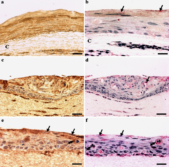
Figure 3.
Expression of P2X5 and P2X7 receptors in paraffin sections of raft cultures of normal human keratinocytes and of CIN 612 (HPV 31) cells. Nuclei were counterstained blue with haematoxylin. (a) P2X5 immunoreactivity (brown) was present throughout all layers of the raft cultures of normal human foreskin keratinocytes, where the staining was confined largely to the cell membranes and the cytoplasm. The raft culture was supported on a collagen matrix (C). Scale bar. 25 μm. (b) P2X7 immunoreactivity (pink) was present in the raft cultures of normal human foreskin keratinocytes, staining weakly within the uppermost layer (arrows). Scale bar. 25 μm. (c) P2X5 immunoreactivity(brown) was present in the CIN 612 (HPV 31) raft keratinocytes, staining all layers of the raft. Scale bar. 50 μm. (d) P2X7 immunoreactivity (pink) was present in the CIN 612 raft and was associated with the cell cytoplasm and nucleus (arrow). Scale bar. 50 μm. (e, f) High power views of CIN 612 (HPV 31) raft cultures: the uppermost layers are highly disorganised, with nucleated cells at the surface of the raft (arrows). There was also positive staining in the cytoplasm of mitotic cells (double arrows) within the raft for both (e) P2X5 receptors (brown) Scale bar 25 μm. and (f) P2X7 receptors (pink). Scale bar. 25 μm.

