
Figure 4.
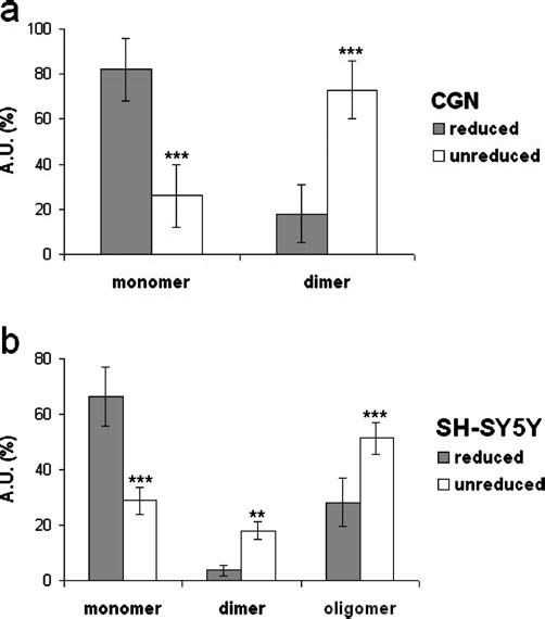
P2Y4 protein expression under reducing and nonreducing conditions. Total protein from cerebellar granule neurons (CGN) (a), and Myc-precipitated proteins recovered from Myc-P2Y4-transfected SH-SY5Y cells (b) were run on sodium dodecyl sulphate polyacrylamide gel electrophoresis (SDS-PAGE) in the absence or presence of 5% β-mercaptoethanol (β-ME) and 50 mM dithiothreitol (DTT). After immunoblotting with anti-P2Y4 or anti-c-Myc (9E10) antibodies, band intensity [expressed as arbitrary units (A.U.)] was analysed by Kodak 1.D Image Analysis software and reported as percent of total protein expression. Data represent means ± standard error of mean (S.E.M) from three (a) and five (b) independent experiments. Statistical analysis was performed using a two-way ANOVA followed by a post-hoc (Newman-Keuls) test for multiple comparisons (*p < 0.05, **p < 0.01, ***p < 0.001)