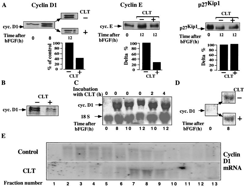
Figure 6.
CLT abrogates expression of cyclins. (A) Quiescent NIH 3T3 cells were stimulated with bFGF and challenged with CLT (10 μM) after 8 hr. Cells were lysed 4 hr later, and 25 μg protein was immunoblotted with antibodies to cyclin D1, cyclin E, or p27Kip1. Note that the upper band in cyclin D1 immunoblot is a different immunoreactive protein because it is not recognized by another anticyclin D1 antibody (see Fig. 8B) and its intensity remains unchanged in serum-starved cells, which do not express cyclin D1. (B) NIH 3T3 cells growing exponentially in bFGF were labeled with 35S-Met-Cys (100 μCi/ml) for 1 hr with or without CLT (10 μM). One hundred migrograms of protein was immunoprecipitated with anti cyclin D1 antibody. Immuncomplexes were separated by SDS/PAGE and visualized by PhosphoroImager. (C) Quiescent cells were stimulated with bFGF for 8 hr, then CLT (10 μM) was added, and cells were harvested either 2 or 4 hr later for Northern blot analysis of cyclin D1 or 18S mRNA. (D) Quiescent NIH 3T3 cells were stimulated with bFGF and simultaneously challenged with or without CLT (10 μM); expression of cyclin D1 mRNA was determined by Northern blotting after 8 hr. (E) RNA extracted from the fractionated sucrose gradients shown in Fig. 2, was separated by formaldhyde-agarose gel electrophoresis and hybridized to cyclin D1 specific probe.

