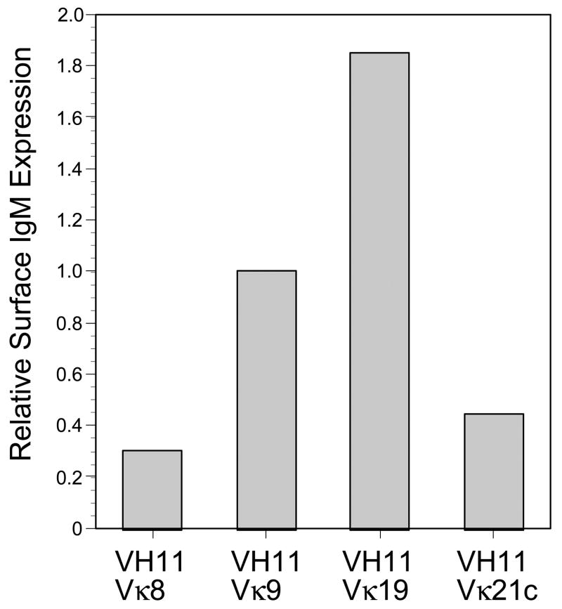
Figure 4.
Cell surface IgM expression on pre-B cells transduced with VH11 and several different light chains. Transduced cells were stained with APC-anti-IgM(331.12) and then gated for VH11 (CFP+) and kappa light chain (GFP+) expression. Fluorescence intensity determined by flow cytometry, normalized for transduction level variation as described in the text. Level on Vκ9 cells arbitrarily set to 1.0.

