Fig. 3.

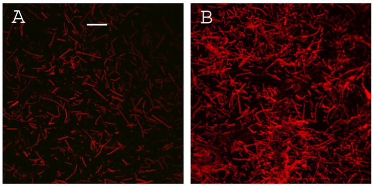
Confocal image of myofibrils contributing to the fluorescence of Fig. 1. (A) – myofibrils on glass coverslip; (B) – myofibrils on glass covered with SIF. Bar is 10 μm. Exposure is the same in each panel.

Confocal image of myofibrils contributing to the fluorescence of Fig. 1. (A) – myofibrils on glass coverslip; (B) – myofibrils on glass covered with SIF. Bar is 10 μm. Exposure is the same in each panel.