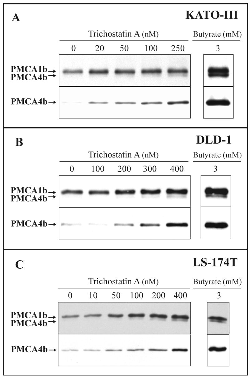
Fig. 7.
Trichostatin A-induced modulation of PMCA expression in various gastric/colon cancer cell lines: concentration dependence.
KATO-III (A), DLD-1 (B) and LS-174T (C) cells were treated with various concentrations of trichostatin A or 3 mM Na+-butyrate as indicated on the panels. Total cellular lysates were prepared from all treatments at day 3 for KATO-III, at day 4 for DLD-1 and at day 2 for LS-174T cells. Equal amounts of cellular proteins (from 20 to 30 μg/lane, depending on cell types and the antibody used for immunostaining) were analyzed for overall PMCA (5F10) and PMCA4b (JA3) expressions.
Treatment of the three cancer cell lines with the HDAC inhibitor trichostatin A led to a marked up-regulation of PMCA4b expression.

