
Figure 5.
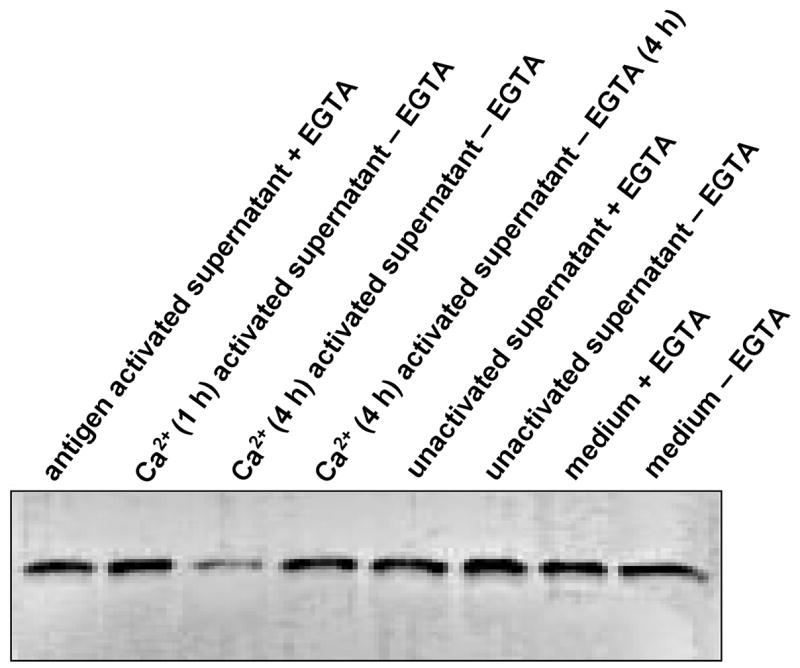
Degradation of MBP by calpain released from activated human MBP-specific T cells. Incubation conditions are described in text. The activated MBP-specific T cells were incubated in the presence of exogenous MBP and with other factors as follows: lane 1, activated MBP-specific T cells + MBP + EGTA (4 h); lane 2, as lane 1 with Ca2+ for 1h but without EGTA; lane 3, as lane 2 for 4 h; lane 4, as lane 3 + EGTA for 4 h; lanes 5–8: unactivated MBP-specific T cells in the presence of exogenous MBP and other factors. Conditions for incubation in lanes 5–8 are the same as lanes 1–4. The MBP degradation was analyzed by SDS-PAGE and immunoblotting.