
FIGURE 10. Genetic deletion of iPLA2β partially, but significantly, potentiates Ang II-induced VASP and Akt phosphorylation in a time-dependent manner.
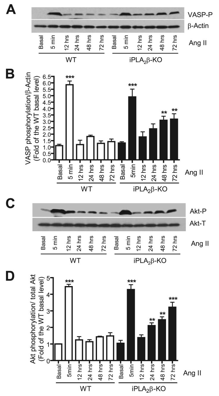
Quiescent mouse WT and iPLA2β-null VSMC were stimulated with Ang II (100 nm) for the indicated intervals. VASP phosphorylation levels (VASP-P, A and B) were determined with anti-VASP Ser-157 phospho-specific antibody and normalized to β-actin and then to the WT basal level. Akt phosphorylation levels (C and D) were determined with an anti-Akt Ser-473 phospho-specific antibody. The phosphorylation level was normalized to the total Akt (Akt-T) level and then to the WT basal level. The representative Western blots (A and C) are from at least three independent experiments and quantitative data are summarized in B and D. **, p < 0.01; ***, p < 0.001 versus WT basal. KO, knock-out.