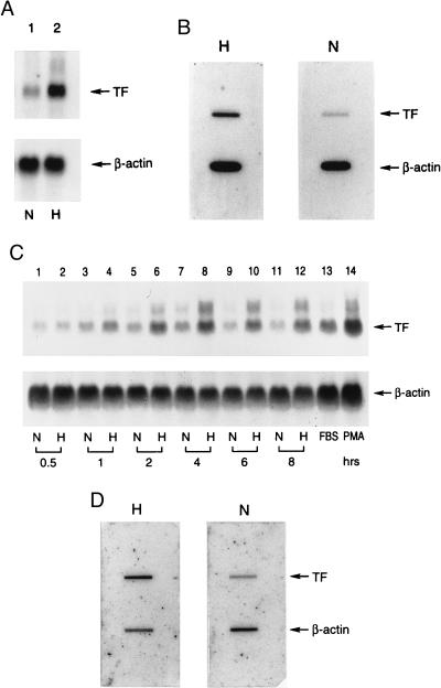
Figure 1.
Effect of hypoxia on tissue factor gene expression in mononuclear phagocytes (A and B) and HeLa cells (C and D). (A and C) Northern analysis for tissue factor transcripts. Human peripheral blood mononuclear phagocytes (≈106 cells, A) or HeLa cells (≈0.5 × 10⁶ cells, C) in serum-free medium were subjected to normoxia (N) or hypoxia (H; pO2 ≈ 12–14 torr) for the indicated times (HeLa) or for 4 hr (mononuclear phagocytes). Northern analysis was performed by loading 30 μg/lane of total RNA and using 32P-labeled cDNA for human tissue factor (Upper) or human β-actin (Lower). FBS and PMA denote cultures exposed to fetal bovine serum (20%) or phorbol myristate acetate (50 ng/ml), respectively, for 1 hr in each case. (B and D) Nuclear run-on analysis for the rate of tissue factor transcription. Mononuclear phagocytes (≈107 cells, B) or HeLa cells (≈106 cells, D) were subjected to normoxia or hypoxia (as above) for 4 hr, nuclei were harvested, labeled by incubation with [α-32P]dUTP for 1 hr, the RNA was isolated, and the same amount of radioactivity was hybridized with cDNA probes for tissue factor or β-actin, the latter already immobilized on nitrocellulose membranes. Results shown are representative of a minimum of four experiments.

