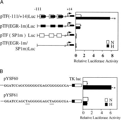
Figure 3.
Hypoxia-inducible tissue factor expression results from transcriptional activation at Egr-1 sites. (A) Transient cotransfection of HeLa cells was performed by using either pTF(−111/+14)Luc, pTF(EGR-1 m)Luc, pTF(SP1 m)Luc, or pTF(EGR-1 m/SP1 m)Luc, and pCMV-β-galactosidase. Cultures were transfected with each of the indicated constructs by using the lipofectamine procedure (GIBCO), and then cells were exposed to normoxia (N) or hypoxia (H) for 5 hr. Luciferase and β-galactosidase activity were then determined. Relative luciferase activity is luciferase activity normalized for β-galactosidase activity. (B) Transient transfection of HeLa cells using pYSF60 (consensus Egr wild-type sequence) or pYSF61 (mutationally inactivated Egr sequence) and pCMV-β-galactosidase by the same procedure described above. Results shown are representative of a minimum of four experiments.

