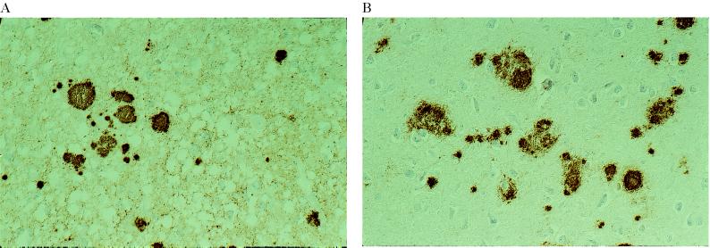
Figure 5.
PrP immunoreactivity in two GSS P102L subjects showing distinct patterns of PrP-res on immunoblot. (A) Cerebral cortex of patient 5. A diffuse, synaptic staining is seen in association with multiple PrP immunopositive plaques. Immunolabeling with 3F4, ×250. (B) Cerebral cortex of patient 6. There are multiple PrP immunopositive plaques. No synaptic staining is visible. Immunolabeling with 3F4, ×250.

