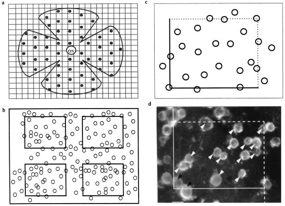
Figure 1.
(a) Systematic random sampling of the retinal surface for stereological counting. ON, optic nerve. (b) Each sample was subdivided into four counting fields after focusing and digitization. (c) Counting was performed by using a stereological dissector. (d) Example of a counted sample from a 5-week-old rd mouse retina cocultured for 8 days with dissociated cells from 8-day-old C57 rod-containing retina. White arrow head, counted cells; black arrow head, excluded cells. (Bar = 5 μm.)

