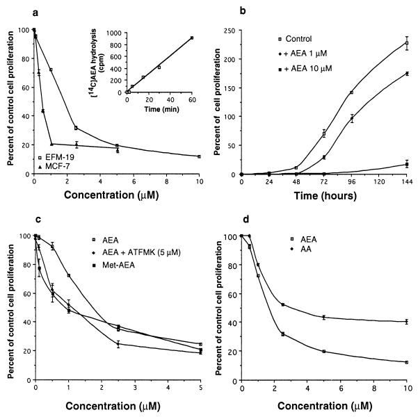
Figure 1.
Effect of anandamide (AEA) on HBC cell proliferation. (a) Dose-dependent inhibition of EFM-19 and MCF-7 cell proliferation by anandamide; the time-dependent hydrolysis of [14C]anandamide by EFM-19 cells is shown in the Inset. (b) Effect of 1 and 10 μM anandamide on the growing curve of EFM-19 cells. (c) Dose-dependent effects of anandamide, with or without the fatty acid amide hydrolase inhibitor arachidonoyl-trifluoromethyl-ketone (ATFMK, 5 μM, Biomol) and of (R)-methanandamide (Met-AEA, Biomol) on EFM-19 cell proliferation; (d) Dose-dependent effects of anandamide and arachidonic acid (AA, Sigma) on EFM-19 cell proliferation. The AEA profiles in a, c, and d are from three different experiments conducted in triplicates. Data in a, c, and d are means ± SEM (n ≥ 3) and are expressed as percentages of control cell proliferation (1 − [Control cell number − treated cell number]/[Control cell number − initial cell number] × 100; 100% = no effect, 0% = maximal cytostatic effect). Data in b are means ± SEM (n = 3) and are expressed as percentages of cell proliferation ([Cell number − initial cell number]/initial cell number × 100). AEA also inhibited [3H]thymidine incorporation into EFM-19 and MCF-7 cell DNA (IC50s were 0.65 and 0.70 μM, and maximal inhibition at 1 μM was 75.0 ± 0.6 and 67.0 ± 0.9%, respectively, mean ± SD, n = 3).

