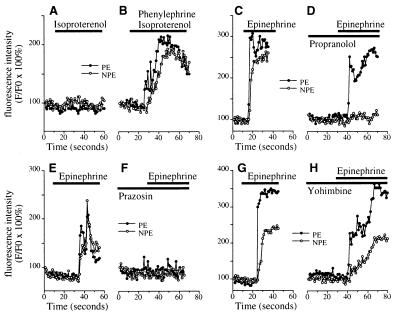
Figure 2.
Pharmacology of adrenergic signaling in the ciliary bilayer. (A and B) The β-adrenergic agonist isoproterenol (100 μM) alone (A) does not increase [Ca2+]i in either the NPE or PE, but together with 100 μM phenylephrine (B) increases [Ca2+]i in both cell layers. Results are representative of the pattern seen in five consecutive experiments. (C and D) Epinephrine (100 μM)-induced [Ca2+]i signaling in the NPE but not the PE is blocked by the β-adrenergic antagonist propranolol (100 μM). Results shown are typical of those seen in five consecutive experiments. (E and F) Epinephrine (100 μM)-induced [Ca2+]i signaling in both layers is blocked by the α1-adrenergic antagonist prazosin (50 μM). Results shown are typical of those seen in five consecutive experiments. (G and H) The α2-adrenergic antagonist yohimbine (100 μM) does not block [Ca2+]i signaling in cells in the ciliary bilayer stimulated with 100 μM epinephrine. Results shown are typical of those seen in five consecutive experiments.

