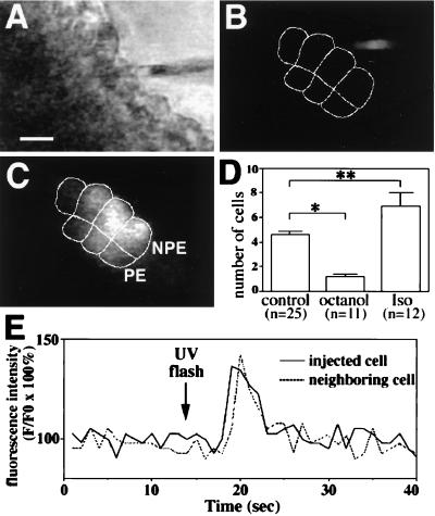
Figure 6.
Intercellular communication in the ciliary bilayer. (A–C) Intercellular spread of lucifer yellow (LY). (A) Transmission image shows the bilayer and the microinjection pipette loaded with LY. (Bar, 10 μm.) (B and C) Serial confocal images show fluorescence in the same field immediately before and 30 s after injection of one of the cells with LY. The micropipette tip can be seen immediately before injection (B), whereas the dye labels four NPE cells and three PE cells (outlined) 30 s afterward (C). (D) Octanol (1 mM) blocks and isoproterenol (100 μM) facilitates cell-to-cell spread of LY. Values are means ± SEM (∗, P < 0.005; ∗∗, P < 0.05). (E) IP3-mediated increases in [Ca2+]i spread from cell to cell in the ciliary bilayer. Caged IP3 was injected into a cell in the bilayer and then uncaged by flash photolysis. A transient increase in [Ca2+]i occurs first in the injected cell, then in a neighboring cell 1 s later. Results are typical of those seen in three separate experiments.

