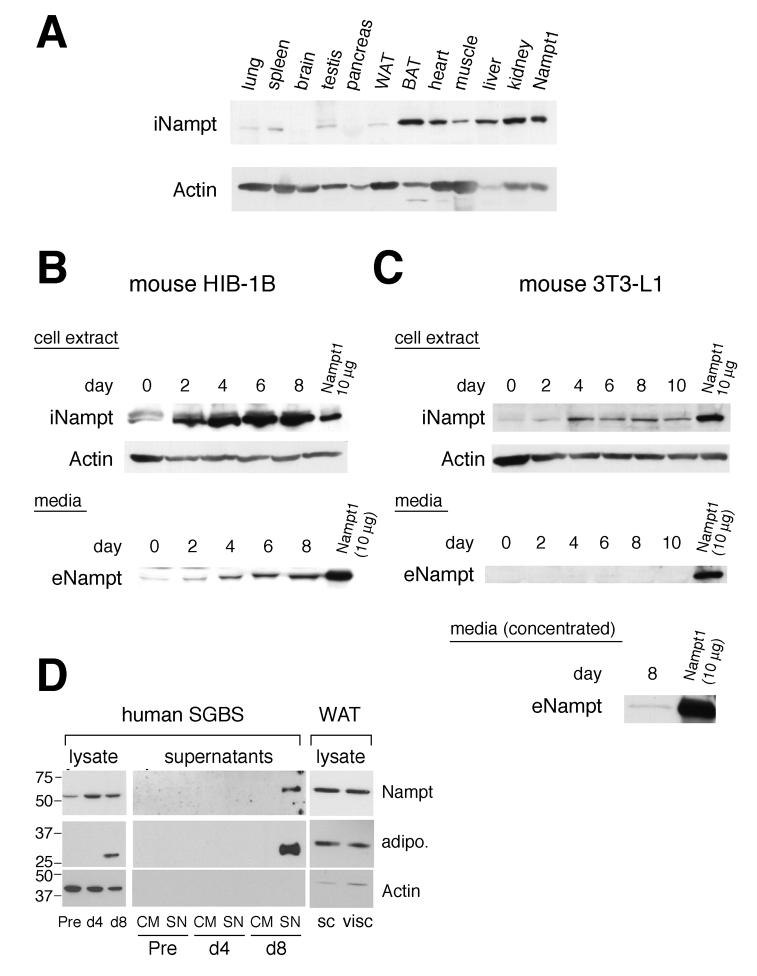
Figure 1.
Tissue distribution of intracellular Nampt in mice and production of intra- and extracellular Nampt during adipocyte differentiation. A) Distribution of intracellular Nampt (iNampt) in mouse tissues. 22.5 μg of each tissue extract from a C57BL/6 mouse was analyzed by Western blotting with Nampt- and actin-specific antibodies. 5 μg of cell extract from a Nampt-overexpressing NIH3T3 cell line (Nampt1) was loaded as a reference. WAT, white adipose tissue; BAT, brown adipose tissue. B) Production of intra- and extracellular Nampt (iNampt and eNampt) during differentiation of HIB-1B brown preadipocytes. Upper panel: Confluent cultures of HIB-1B cells were differentiated, and cell extracts were prepared at the indicated days. 45 μg of each cell extract was analyzed. Lower panel: 20 μl of each culture supernatant collected at the indicated days was analyzed. C) Production of iNampt and eNampt during differentiation of 3T3-L1 white preadipocytes. Upper panel: 45 μg of each cell extract collected at the indicated days was analyzed. Middle panel: 20 μl of culture supernatant was analyzed. Lower panel: Concentrated culture supernatant was analyzed at day 8 to detect eNampt. 10 μg of the cell extract from Nampt-overexpressing fibroblasts (Nampt1) was loaded as a reference in each experiment. D) Production of iNampt and eNampt during differentiation of human SGBS white preadipocytes. Left and middle panels: 13 μg of each cell extract and 25 μl of each culture supernatant (10-fold concentrated) collected at the indicated points were analyzed. Adiponectin production is also shown as a positive control for adipocyte differentiation. Pre, undifferentiated preadipocytes; d4, differentiating adipocytes at day 4; d8, mature adipocytes; CM, control medium; SN, supernatant. Right panel: iNampt protein expression was analyzed in tissue extracts from human subcutaneous (sc) and visceral (visc) white adipose tissues (WAT).

