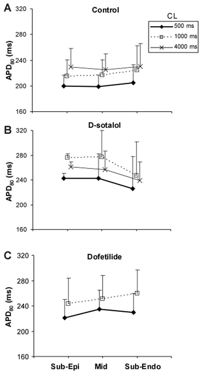
Figure 4.

Intramural APD distribution. Panel A, APD80 measured in sub-epicardial (Sub-Epi), mid-myocardial (Mid) and sub-endocardial (Sub-Endo) muscle layers at pacing CL of 0.5, 1 and 4 s in control conditions. The data were obtained from 5 hearts perfused with either Tyrode solution or blood. Panels B and C, APD80 in different tissue layers in the presence of 125 μM of d-sotalol (B) and 2 μM of dofetilide (C).
