Figure 4.
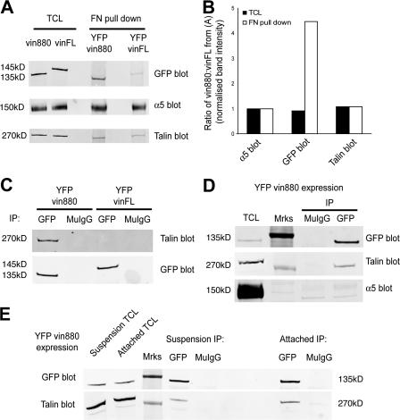
YFP-vin880 is enriched in FN– integrin complexes and coimmunoprecipitates talin and integrin. (A) FN–bead bound complexes isolated from NIH3T3 cells expressing either YFP-vin880 or -vinFL and immunoblotted for GFP, α5 integrin, or talin. (B) Quantification of A expressed as a ratio of vin880/vinFL after immunoblot band signal intensity normalization to the respective α5 integrin signal intensity. Open bars represent ratios of indicated proteins from pull-down experiments with pFN-coated beads; shaded bars represent protein ratios detected in total cell lysate (TCL) of cells expressing YFP-vin880 or -vinFL. (C) Immunoprecipitations using anti-GFP or control mouse IgG (MuIgG) from NIH3T3 cells expressing either YFP-vin880 or -vinFL. (D and E) Immunoprecipitations using anti-GFP or control MuIgG from NIH3T3 cells expressing YFP-vin880 after treatment with a chemical cross-linker (D) or from cells in suspension for 15 min (E) versus cells left attached to tissue culture dishes. All blots are representative of more than two independent transfections. Mrks denotes the position of molecular mass standards (250 kD for talin blots and 150 kD for GFP and α5 integrin blots), which are visible by Western blotting using the infrared imaging system.

