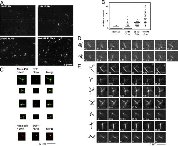
Figure 6.
Orthogonal branching of F-actin by FLNa. (A) Relationship between FLNa concentration and filament branching. Treatment of actin filaments tethered to a surface by gelsolin with FLNa and free F-actin results in the formation of orthogonal actin filament crossings and branches. An increase in filament junctions is observed with the addition of increasing FLNa concentrations. (B) Plot of branch number versus FLNa concentration added. For each experiment, junctions in 40 randomly selected areas in >5 coverslips (3 experiments) were analyzed. (C) Fluorescence microscopy of F-actin-FLNa junctions. The presence of FLNa at the junctions is detected using RFP or EGFP labeled FLNa and Alexa 488- or Alexa 568 phalloidin-labeled F-actin, respectively. (D) Representative time-lapse images that record the movement of actin filaments bound to anti-gelsolin IgGs (see Video 1). Note that the gelsolin-tethered end of the filament is immobile while the free end oscillates rapidly. The superimposed tracings to the left track the movement of the filament with time. (E) Behavior of actin filaments cross-linked by FLNa molecules. FLNa creates stable orthogonal filament cross-links that are not deflected by thermal motion. Filament junctions are at nearly perpendicular angles. Note that the addition of filament stabilizes it in the vicinity of the cross-link. The images on the left record the filament movements (see Video 2).

