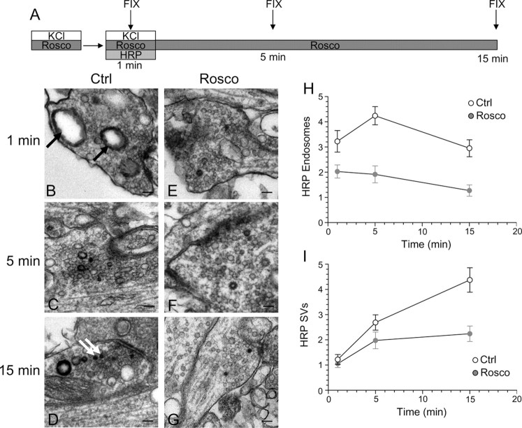
Figure 8.
Inhibition of cdk5 selectively inhibits endosome generation and not single SV endocytosis. A, Cultures were stimulated for 10 s with 50 mm KCl, repolarized for 10 min, and then stimulated again with 50 mm KCl for 2 min in the presence of HRP. Cultures were repolarized for up to 15 min in the absence of HRP. Cultures were fixed at regular intervals during this time course and were preincubated with 50 μm roscovitine (Rosco) for 10 min before the first KCl pulse and at all steps up to fixation where indicated. B–G, Representative electron micrographs of HRP uptake in control (B–D) or roscovitine-treated (E–G) nerve terminals at 1, 5, and 15 min after stimulation. Black arrows indicate HRP-labeled endosomal structures whereas white arrows indicate HRP-labeled SVs. Scale bars: 200 nm. H, Mean number of HRP-labeled endosomes per nerve terminal. I, Mean number of HRP-labeled SVs per nerve terminal. In H and I, open circles represent control and closed circles represent roscovitine (Ctrl: 1 min, n = 99 nerve terminals; 5 min, n = 213; 15 min, n = 107; Rosco: 1 min, n = 100; 5 min, n = 68; 15 min, n = 43). Error bars indicate SEM.

